The Society met in the Ebenezer Duncan Centre at The Victoria Infirmary on Thursday 24th November 2005 at 7pm. The President, Dr Philip Wilson, was in the chair.
Sederunt 35
I Apologies
Apologies were received from Dr David Kidd.
II Minutes
The minutes of the meeting of 13th October were accepted. Dr Wilson reminded members that minutes are published on the Society's website in addition to the written minute in the Society's minute book.
III Christie Cup
Dr Wilson presented the Christie Cup to Dr Ewing Forrester for winning the Bogey Competition at the Society's golf outing on 6th September.
IV Next meeting
Dr Wilson announced that the next meeting of the Society will be a joint meeting with the Royal Medico-Chirurgical Society on Thursday 12th January 2006 at the Royal College of Physicians and Surgeons of Glasgow at 7.00 for 7.30pm. The lecture will be given by Prof Alan Silman, ARC Professor of Rheumatic Disease Epidemiology and Director of the ARC Epidemiology Unit at the University of Manchester. His talk will be entitled 'Clinical trials – do they always give the right answer?'
V Dr Willox's lecture
The President then invited Dr David Willox to give his lecture to the Society recounting his experiences as medical officer for the charity Glasgow the Caring City in the aftermath of the Asian tsunami. A précis of the paper follows this minute.
At the close of Dr Willox's talk, and following interesting discussion, the President called upon Dr Duncan Macintyre to move a vote of thanks to Dr Willox. This was most heartily responded to by members of the Society.
This was all the business and Dr Wilson closed the meeting.
Tsunami – after the wave
Dr Willox described Sri Lanka as a beautiful land populated by beautiful people. The warm currents of the Indian Ocean lap on sandy beaches. Along the coast, fishermen and their families live in small villages beside the shore. You might be forgiven for thinking that this is paradise. This is the picture you might have imagined on 25th December, 2004. Within 24 hours, all this changed. The Asian tsunami on Boxing Day killed a quarter of a million people, of whom 40,000 died in Sri Lanka.
As soon as reports of the tsunami reached the UK, local charity Glasgow the Caring City mobilised support and began collecting donations. Through the involvement of his wife Morag, Dr Willox volunteered to drive a van round Glasgow to uplift bags of clothes and other gifts. He soon found himself acting as medical officer to help sift through donations of medical items. It was a natural next step to join the small team of volunteers and fly out to Sri Lanka to begin the long process of supporting relief and regeneration.
Dr Willox described the destructive power of the waves. He showed photographs of shattered buildings, dead animals and birds, and the massive human toll. He showed how the scale of the debris was overwhelming. On closer inspection, what appeared to be small mounds of wood and masonry were, in fact, the bodies of men, women and children. In temperatures of 42°C and oppressive humidity, the smell was unbearable.
In March 2005, three months after the tsunami struck the coast of Sri Lanka, a team of volunteers from Glasgow the Caring City flew out to the Hikkaduwa region. Dr Willox and the rest of the team spent two weeks assessing needs and setting up projects. The only forensic pathologist in the area, Dr Clifford Perera, had been faced with the task of managing 6500 bodies. To put that in perspective, relief workers from Leicester indicated that 200 bodies over two weeks would be enough to strain local services in the UK. In spite of this, Dr Perera remained remarkably positive and the relief team were able to help him with donations of computer equipment and fund travel to a conference in Thailand on disaster victim identification and management.
After the terrorist attack on the twin towers in New York on September 11th 2001, people posted photographs of their missing relatives on a wall. In contrast, Dr Perera was only able to create a 'wall of the dead' – photographs of the dead bodies yet to be identified. In the scramble to recover and identify bodies, villagers claimed that a team of scientists from Interpol investigating the deaths of foreign nationals had shown callous disrespect in their handling of the bodies of local people.
Dr Willox went on to show photographs of the Galle to Colombo train which was partially derailed at Peralyia by the first tsunami wave. As the water withdrew, people sought refuge in the railway carriages, only to be caught by the second wave. There were stories of people lying injured and dying three days later when the press and news media arrived. Government rescue teams are said to have taken a further seven days to reach the scene. The official death toll was given as 1500, though the true figure is certainly much greater. In spite of this, Dr Willox was reluctant to criticise the Sri Lankan government given the scale of the disaster. A permanent memorial of three railway carriages has been created on the site.
It is easy to underestimate the difficulty of conducting worthwhile work in the aftermath of such a large disaster. The Disasters Emergency Committee had received donations amounting to £300 million, but the Sri Lankan government had been slow to identify priorties. In Peralyia there was an urgent need to provide shelter before the arrival of the monsoon season. Charities had built terraces of wooden huts. Unfortunately the local practice of using open fires for cooking had led to some shelters catching fire and being destroyed. Dr Willox alluded to conflicts of interest between fishermen who lived beside the sea for access to their boats, and corporate plans to build luxury hotels on the beaches. The 100 metre rule instituted after the tsunami prevented villagers from rebuilding their dwellings within 100m of the high tide mark. A model village had been built 10 kilometres inland, an impractical proposition for the fishermen. Other examples of poor relief work included the rebuilding of an obstetric ward in a hospital run by Dr Weerasinghe in Arachikanda at a time when delivery rates were falling as more women were going to a local consultant unit. Dr Weerasinghe dealt with up to 150 patients a day with very limited resources, and it was felt that the money might have been better spent.
On a smaller scale, there were some valuable charitable projects. With the rainy season about to start, the medical officers were concerned about the spread of infectious disease, particularly Dengue Fever and Japanese B Encephalitis. The tsunami had destroyed refrigerators in local hospitals resulting in the inability to store vaccines. This meant that children were no longer being immunized against infectious diseases. Glasgow the Caring City was able to help by donating fridges.
The library at Peralyia had survived the flood and had been transformed into a makeshift medical centre. Instrumental in this was a remarkable American nurse, Alison Thompson. Also here were Dr Shouren Datta and Dr Carolyn Datta who had been working in Chennai. On receiving news of the tsunami, they had moved to Sri Lanka to help with the relief work. Both were well known to members of the Society as Dr Shouren Datta had worked in gastroenterology at the Victoria Infirmary.
A principal aim of the charity was to engage in projects that were sustainable. An 82 year old rice farmer had had his paddy fields inundated by salt water. The charity was able to supply him with rice seed for replanting once the monsoon had washed the salt from the soil. This will enable him to feed himself and five other families. The Raka Institutional Complex provided respite care for the victims of abuse. With help to improve the rooms and by training more teachers it would be able to cater for an influx of 800 orphans.
Dr Willox accepted that much of the work amounted to applying a sticking plaster on a gaping wound, but through small acts, Glasgow the Caring City was able to make a material difference to the lives of ordinary people. He spoke briefly of some future plans to arrange surgery for two deaf twin boys. Dr Willox encouraged members of the Society to support the work of Glasgow the Caring City as it continues to provide help in Sri Lanka and other areas of the World.
Listen again:
Download hi-fi mp3 78.0Mb
Download lo-fi mp3 39.0Mb
Listen to lo-fi stream
Further reading:
Glasgow the Caring City
Sri Lanka Report
After the Tsunami: Legal Implications of Mass Burials of Unidentified Victims in Sri Lanka
Revisiting the Tsunami: Health Consequences of Flooding
Alison Thompson's diary
The Waste Land
TS Eliot
Maslow's hierarchy of needs
tags: tsunami sri lanka
25 November, 2005
16 November, 2005
100 years ago: Session 1905-06; Meeting IV - Sleeping Sickness
Sederunt 35
The Society met in the rooms of the Medical Club 22 Carlton Place on Thursday 16th November 1905 at 9 p.m.
The President, Professor Stockman, was in the chair, and in all 35 gentlemen were present.
I Minutes
The minutes of last meeting were read and approved.
II New Members
1. On the motion of the Chairman the ballot was dispensed with and Dr Edward J. Primrose was declared elected a member of the Society.
2. The Secretary read two proposals for membership viz.
Dr Wm. Barr Inglis Pollock 13 Belgrave Terrace
Proposed by Dr Alexander Morton.
Seconded by Dr John P. Duncan.
Dr John Paton, 21 Moray Place
Proposed by Dr James Hamilton.
Seconded by Dr James Weir.
III Captain Greig's paper
Captain Greig I.M.S. then gave a most interesting & full account of the researches on "Sleeping Sickness", conducted by the Royal Society's Commission in Uganda and the Nile Valley, and showed many lantern slides illustrative of his remarks.
A precis of the paper follows this minute.
At the close of Captain Greig's account the President moved a hearty vote of thanks to Captain Greig & this was at once heartily responded to. Professor Stockman also made some remarks on paracytic diseases carried by means of flies, and on the action of arsenic in improving and alleviating the condition of the patient in many diseases without actually bringing about a permanent cure.
Professor Muir then made a few remarks anent the absence of immunity even after the long course pursued by the disease; & in this connection said he thought innoculation would probably be useless. He then referred to Ehrlich's work on the aniline dyes and their action on paracytic diseases.
Drs McGilvray, Reid, Halliday, Burgess & Dunlop also took part in the discussion & asked various questions.
Captain Greig then replied to the questions and explained some points he had omitted to refer to in his account.
This was all the business.
Precis of Captain Greig's paper
The serious nature of the disease was first referred to. It is estimated that there have been 100,000 deaths from it since the outbreak of the epidemic in 1900, and there is the possibility of it spreading widely along trade routes. The geographical distribution of the disease was then spoken of. In the Uganda district it is found mainly on the shore and islands at the northern end of Victoria nyanza & affects a belt measuring from 10 to 15 miles from the shore. Until 5 or 6 years ago this was practically a closed district but since then caravans have opened it up & no doubt these serve to carry infection along the trade routes.
The symptomatology of the disease was next discussed. It may be divided into 3 stages, 1st onset with few or no symptoms 2nd stage of sleepiness & emaciation 3rd stage of sleep ending in death with extreme emaciation.
Temperature
Stage 1. may be normal for 2 or 3 years.
Stage 2. Slight irregularity with slight fever.
Stage 3. Markedly subnormal for a week or two before death & may fall to the temperature of the surrounding air viz about 93° F.
Enlarged glands are present all over the body in the 2nd stage, & these are full of the paracyte of the disease.
The sleepy look appears on the face during the 2nd stage.
Emaciation always appears during the second stage & sometimes marked nervous symptoms also come on e.g. acute mania.
Profound sleep, from which however the patient may be roused by vigorous measures appears in the third stage & this is accompanied by extreme emaciation & always ends in death.
The blood contains the paracyte in the early stages of the disease and it is also present in the cerebrospinal fluid in the later stages.
Pathology of the disease.
The lymph glands both superficial & deep are enlarged & full of the paracyte. The Brain presents no special features macroscopically but microscopically there are crowds of leucocytes round the vessels & in the last stages streptococci are present. In both blood & cerebrospinal fluid there is a marked increase of mono-nucleated white cells. The stomach presents numerous small ulcers due to the digestion of submucous petechiae and in the heart numerous petechiae are also present.
Experimental work was carried out in a woodlined-iron laboratory. Monkeys were largely used for experiment.
Characteristics of the Paracyte.
It is a trypanosome & belongs to the flagellata. It reproduces itself by fission & sometimes it may divide more than once without separation of the new individuals from each other so that a rosette is formed.
It is an elongated fusiform body possessing nucleus, micro nucleus, a thin vibratory fin extending along its whole length and terminating in a simple flagellum the micro nucleus being at the opposite pole to the attachment of the flagellum. Vacuoles & what seem to be food particles are usually present & the organism is about 3 times the length of the diametre of a red blood corpuscle. Similar forms of trypanosomata are also found in various forms of cattle disease in Uganda & Abyssinia.
Large numbers of natives in the infected area and also in non infected areas were examined. The blood cerebrospinal fluid & gland juices were investigated. The details of the methods adopted in such examinations were discussed. It was found that from 50 to 70 % of the population in the affected area was infected & in the early stage of the disease, while in a large number of diseased & healthy natives examined in non infected areas no trypanosomata were found. A number of the infected cases were followed out. Some died in the 3rd Stage: some died of pneumonia: some are fairly well still, & some may recover, but no case of recovery from the 3rd Stage is known. The exact area of distribution of sleeping sickness was mapped out and in these areas a fly (Glossina Palpalis) was found to be abundant, while in the free areas it was absent. Imported cases dying in a fly-free area do not spread the disease for other biting flies cannot carry the paracyte & infect man. If one Glossina Palpalis bite an infected subject and take up from his blood a single Trypanosome the paracyte proliferates enormously in the stomach & the next man punctured by the fly's proboscis becomes infected. This fly does not flourish in the open but prefers the cooler & shadier forrest. Many of these flies were captured & after being fed on an infected subject were caused to bite a healthy monkey at varying periods after ingestion of the infective meal. It was found that 48 hours seemed to be the limit of infectivity of a fly after infection, but that the monkey bitten within that period always developed the disease. Flies freshly caught in the sleeping sickness area were proved to be infective.
Treatment.
Arsenic & aniline dyes in combination were used & have an effect in reducing the numbers of paracytes present in the body & no doubt prolong life, but relapses constantly occur. This part of the investigation is still being vigorously worked at. Many interesting slides were then shown to illustrate the type of country and mode of life there of natives & whites & finally it was explained with reference to animal trypansomiasis that wild animals, themselves immune, might contain in their blood thousands of trypanosomata and thus act as reservoirs of the disease.
In reply to the remarks made by various gentlemen it was explained that no doubt the greater care exercised by the whites, their greater resistance, and the wearing of clothes, were factors in the prevention of the disease in their case; but one white man, a gardener, had been infected & is at present in this country ill with the disease. The incubation period seems to be from 8 days to 3 weeks. Wild monkeys do not permit the fly to bite them & so escape the disease.
Ralph Stockman
Archive: Royal College of Physicians and Surgeons of Glasgow
Reference: GB 250 RCPSG 73/1/11 Minute Book No. 6
Further reading
Paul Ehrlich
Uganda
Nyanza
Abyssinia
Sleeping sickness
Glossina palpalis
14 November, 2005
Notice of meeting: 'Tsunami - after the wave'
You are cordially invited to attend the meeting:
'Tsunami - after the wave'
Medical charity work in Sri Lanka
Speaker: Dr David Willox
Southside GP
Date and time: Thursday 24th November 2005 at 7.00pm
Venue: Ebenezer Duncan Centre, The Victoria Infirmary, Glasgow
This meeting is open to all medical, nursing and paramedical staff.
If you are a member of the Society and you would like to book for the pre-meeting buffet, please email 'Buffet' and your name to: Lesley.O'Donnell@gvic.scot.nhs.uk
Please note that the lecture and subsequent discussion will be recorded for later publication on the Society's website.
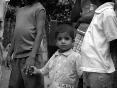
Photo credit: oregongirl! published here under a non-commercial Creative Commons licence.
02 November, 2005
100 years ago: Session 1905-06; Meeting III - The use of the Cystoscope
Sederunt 34
The Society met in the rooms of the Medical Club, 22 Carlton Place on the evening of Thursday 2nd November 1905 at 9pm.
The President, Professor Stockman, was in the chair and in all 34 gentlemen were present.
I Minutes
The minutes of the last two meetings were read and approved of.
II Correspondence
The secretary read a letter from the Medico-Chirurgical Society, Glasgow, in which members of the Society were invited to be present at an address, to be given, on Nov. 3rd by Mr W. Sampson Handley F.R.C.S. & Hunterian Professor, Royal College of Surgeons England, Subject: "On the mode of spread of Breast Cancer: with special reference to operative treatment."
III New Members
The secretary read a proposal for membership viz.
Dr Edward J. Primrose 551 Dumbarton Rd. Partick
Proposed by John P. Duncan
Seconded by R. Wardrop Forrest
IV Dr Newman's demonstration
Dr David Newman then gave a most interesting and instructive demonstration on "The use of the Cystosope" & illustrated his remarks by means of an opaque projector.
A precis of the demonstration follows this minute. The Chairman at the conclusion of the demonstration made a few remarks on the subject and moved a vote of thanks to Dr Newman. This was most cordially responded to, and then Dr Newman replied in a few words.
V Tariff of Fees
The Secretary moved as follows:
"That, as recommended by the Council of the Society, the Tariff of Fees printed at the end of the book of laws be adopted by the Society".
He explained that the Tariff had not been readopted for several years, and that it was of some importance, in the case of legal proceedings being taken by a member of the Society for recovery of fees, that the tariff should be formally readopted by the Society from time to time.
This was carried [...]
VI Dr J. C. MacEwen's statement
Dr J. C. MacEwen intimated that he had been requested to ascertain the views of the Society on the proposal brought forward by the Eastern Medical Society that a dance be held this winter under the auspices of the Southern, Eastern & Northern Medical Societies. After some discussion, on the suggestion of Dr McGilvray, it was agreed that the matter came within the province of the Amusements Committee of the Medical Club & that Dr MacEwen should approach this committee.
This was all the Business.
Precis of Dr Newman's Demonstration
Dr David Newman demonstrated by means of an opaque projector the cystoscopic appearances of the urinary bladder as presented by the mucous membrane of the trigone and the orifices of the ureters in certain diseases of the kidney. He described his first electric cystoscope (January 1883) still armed with the first electric lamp introduced into a human bladder, and then showed his present cystoscope, which fulfilled the following requirements:–
1. Comparatively small lumen of stem to avoid injury to the urethra or neck of bladder.
2. Large field of vision and a clear view.
3. Easy means of clearing the [...] , should it become obscured, without [...] stem of instrument from bladder
4. Good illumination without danger of scalding the mucous membrane of the bladder.
5. Ease in sterilizing the instrument
6. Facility in emptying the bladder should it be necessary without removing the cystoscope.
7. Ability to demonstrate to a second observer the object seen
8. Means of steadying the instrument during [...] of fixing prism cystoscope at any point, and of defining clearly position of a lesion.
He then demonstrated, by means of a special opaque projector the appearances seen through the cystoscope, so arranged that only a portion of the diagram was illustrated at one time upon the screen: by moving the diagram the appearance of the mucous membrane was seen, bit by bit, as in an inspection with the cystoscope. He first illustrated some easily recognised lesions of the viscal mucous membrane, such as hyperaemia of the bladder in a case of injury to the medulla oblongata, and then an extensive series of morbid conditions, such as ulcers, encysted and other calculi, new growths, ascending and descending urethritis, shoots of blood from the ureters. The last named condition was demonstrated by means of a most impressive working model.
Finally he drew the following conclusions:-
I. When one ureter orifice is altered, the other normal, the renal lesion is on the side of the [...] ureter.
II. When the urinary shoots are more frequent on one side than on the other
a. greater functional activity is indicated by the shoots being uniform in size and regular in rhythm.
b. undue irritation of the kidney is inferred when the shoots, while frequent are irregular in rhythm, unequal, and small in size.
c. Stricture, stone, or chronic ureteritis is suspected when the shoots are distorted in form or irregular in amount.
III. When the urine does not escape in distinct jets
a. dilatation of ureter without paralysis of sphincter is indicated when the urine dribbles into the bladder at intervals.
b. paralysis of sphincter is shown by urine flowing into bladder almost continuously.
IV. The character of morbid fluids escaping from the ureter, or of clots of blood &c, occupying its opening, denotes the changes taking place in the corresponding kidney.
V. The deformity of the orifice also indicates the character of the renal disease
a. Pin-head contraction (chronic inflammation, or impacted calculus)
b. elongated and distorted (distension of renal pelvis or infected nephritis)
c. swollen or pouting (prolonged but not acute inflammation of the renal parenchyma)
d. dilated (advanced tuberculous or calculous pyonephrosis)
e. U shaped (significance doubtful – usually denotes prolonged irritation of renal pelvis).
Ralph Stockman
Archive: Royal College of Physicians and Surgeons of Glasgow
Reference: GB 250 RCPSG 73/1/11 Minute Book No. 6
Subscribe to:
Posts (Atom)


